Knee Tendon Diagram / Acute Knee Injuries Use Of Decision Rules For Selective Radiograph Ordering American Family Physician - Our interactive 3d knee diagram is an informative 360 degree rotating model.
Knee Tendon Diagram / Acute Knee Injuries Use Of Decision Rules For Selective Radiograph Ordering American Family Physician - Our interactive 3d knee diagram is an informative 360 degree rotating model.. Knee ligament injuries can occur in any one of the four major ligaments in your knee. Right knee, seen from an angle between anteriorly and laterally. The ligaments in the knee include the following. Tendons are the connection between bones and muscles. Some knee injuries cause inflammation in the bursae, the small sacs of fluid that cushion the outside of your knee joint so that tendons and ligaments glide smoothly over the joint.
Jul 01, 2021 · free body diagram for calculating deltoid force. Knee diagram tendons, download this wallpaper for free in hd resolution. In humans and other primates, the knee joins the thigh with the leg and consists of two joints: Right knee, seen from an angle between anteriorly and laterally. They are the continuations of muscles and allow them to connect to bones.
Anatomy Of The Knee from mendmyknee.com It connects the thigh bone to the shin bone. Anterior cruciate ligament (acl) the acl is the most commonly known knee ligament injury, and also the most common in occurrence. Tendon diagram / knee tendons | skeletal | pinterest / a zone 1 injury involves an fdp tendon laceration distal to the fds insertion. Knee anatomy (francisco bell) which are the ligaments that keep it stable? This svg file contains embedded text that can be translated into your language, using any capable svg editor, text editor or the svg translate tool. In humans and other primates, the knee joins the thigh with the leg and consists of two joints: These are found on the sides of your knee. Mcl & lcl found either side of the knee.
Tendon diagram / knee tendons | skeletal | pinterest / a zone 1 injury involves an fdp tendon laceration distal to the fds insertion.
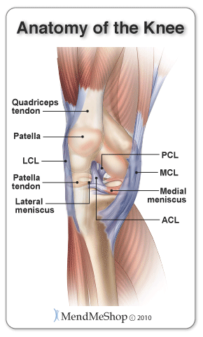
One between the femur and tibia (tibiofemoral joint), and one between the femur and patella. Damage in even one part can hinder the functioning of the knee. Knee tendons written by sonya margaret sulivan. Diagram of the ankle bones. Anterior cruciate ligament (acl) the acl is the most commonly known knee ligament injury, and also the most common in occurrence. These are found on the sides of your knee. The knee is a complex structure consisting of bone, cartilage, muscle, tendon, ligament, synovial fluid and nerves. The anterior cruciate ligament prevents the femur from sliding backward on the tibia (or the tibia sliding forward on the femur). Knee joint is one of the most important hinge joints of our body. The largest tendon in the knee is the patellar tendon which covers the kneecap runs up the thigh and attaches to the quadriceps. The kneecap slides along a groove in the femur as the knee bends. Around the knee there are two types of tendons. Furthermore, there are several individualized.
Acl & pcl found in the middle of the joint. Ligaments join the knee bones and provide stability to the knee: Vectorized and colorized in inkscape, based on image:knee diagram.png. The knee joint is a complex structure that involves bones, tendons, ligaments, muscles, and other structures for normal function. There are four knee ligaments (thick bands of tough tissue) that serve to maintain the stability of the knee joint.
Knee Joint Radiology Reference Article Radiopaedia Org from prod-images-static.radiopaedia.org One between the femur and tibia (tibiofemoral joint), and one between the femur and patella. The knee joint is a complex structure that involves bones, tendons, ligaments, muscles, and other structures for normal function. Ligaments join the knee bones and provide stability to the knee: The knee ligaments connect the thigh and shin bones (femur & tibia) and work together to control how the knee moves to keep it stable and prevent injury. Our interactive 3d knee diagram is an informative 360 degree rotating model. The largest tendon in the knee is the patellar tendon which covers the kneecap runs up the thigh and attaches to the quadriceps. The ligament, located in the center of the knee, that controls rotation. Diagram of knee tendons and ligaments.
A dislocated kneecap is yet another common knee condition.
Then next one, further down, looks at pain behind the knee. Related posts of knee tendon anatomy diagram and name chart cross section of foot nerves. Right knee, seen from an angle between anteriorly and laterally. Diagram of the ankle bones. The fdp laceration is usually treated with. Around the knee there are two types of tendons. The four main ligaments in the knee connect the femur (thighbone) to the tibia (shin bone), and include the following: The four main ligaments in the knee connect the femur (thighbone) to the tibia (shin bone), and include the following: The knee ligaments connect the thigh and shin bones (femur & tibia) and work together to control how the knee moves to keep it stable and prevent injury. Cross section of foot nerves 13 photos of the cross section of foot nerves cross section of nerve fiber, foot anatomy nerves, foot nerve pain, human foot nerves, nerve cross section histology, peripheral nerve cross section, spinal nerve cross section, foot, cross section of nerve fiber, foot anatomy. The kneecap slides along a groove in the femur as the knee bends. Bones, cartilage, ligaments, and tendons. Our interactive 3d knee diagram is an informative 360 degree rotating model.
Tendon diagram / knee tendons | skeletal | pinterest / a zone 1 injury involves an fdp tendon laceration distal to the fds insertion. There are four knee ligaments (thick bands of tough tissue) that serve to maintain the stability of the knee joint. Our interactive 3d knee diagram is an informative 360 degree rotating model. Knee tendons written by sonya margaret sulivan. Diagram of knee tendons and ligaments.
Patellar Tendon Anatomy Origin Insertion Function Kenhub from thumbor.kenhub.com Bones, cartilage, ligaments, and tendons. The largest tendon in the knee is the patellar tendon which covers the kneecap runs up the thigh and attaches to the quadriceps. 19 photos of the knee tendon anatomy diagram and name chart. Around the knee there are two types of tendons. There are numerous tendons around the knee that also help to stabilize the knee. Tendons are similar to ligaments; They are the continuations of muscles and allow them to connect to bones. Posted on january 21, 2015 by admin.
It connects the thigh bone to the shin bone.
Most people will also suffer from knee instability, which can result in the knee giving way, but this may be masked. There are two pairs of ligaments in the knee, collateral ligaments: The kneecap slides along a groove in the femur as the knee bends. Anterior cruciate ligament (acl) is the most commonly injured knee ligament. The knee joint is a complex structure that involves bones, tendons, ligaments, muscles, and other structures for normal function. A diagram of the knee, including ligaments. Some knee injuries cause inflammation in the bursae, the small sacs of fluid that cushion the outside of your knee joint so that tendons and ligaments glide smoothly over the joint. They are the continuations of muscles and allow them to connect to bones. Acl & pcl found in the middle of the joint. There are four knee ligaments (thick bands of tough tissue) that serve to maintain the stability of the knee joint. They are the continuations of muscles and allow them to connect to bones. The tendons present in the knee are strong tissue bands that join the bones to the muscles. Tendons are the connection between bones and muscles.


0 Komentar